The Bigger Picture
How is mitosis adapted to fit the complex native environment of a dividing cell?


Mitosis comprises a series of beautiful, highly dynamic events, carefully orchestrated to ensure the production of two genetically identical daughter cells and must proceed with high fidelity trillions of times over the average human lifetime. Mitosis of adult stem cells maintains tissue homeostasis, particularly in organs with a high rate of cellular turnover, and permits tissue regeneration upon damage. Mitosis can also be pathogenic - cancer is necessarily a disease of cell division, with aberrant mitoses driving tumor expansion and enabling subsequent pathologies.
Real-time observation (live cell imaging) has formed the basis for much of our current understanding of mitosis. Traditional model systems for live imaging of mitosis (e.g. cell culture models) often lack cellular, environmental and physiological complexity. As a result, how fundamental mitotic processes are influenced by the myriad intrinsic and extrinsic factors that form a cell’s normal environment in vivo remains relatively unexplored. Importantly, mitosis in both adult stem cells and tumor cells is likely to be influenced by these types of factors.
(HeLa movie courtesy of J. Ryan)


Embryonic blastomeres

Adult GSCs
Model genetic organisms have proven an invaluable tool in the study of mitosis and Caenorhabditis elegans is uniquely well-suited as a model for investigating mitosis in situ. C. elegans is a small nematode worm, which like other genetically tractable organisms, is suitable for large-scale genetic screens and benefits from a wealth of genetic and cell biological tools. Adult animals exhibit cellular and physiological complexity, with analogous cell types and conserved signaling pathways that serve parallel functions in humans. Unlike other multicellular model organisms, C. elegans adults are small and transparent, making them amenable to live-imaging studies in whole-mounted animals.
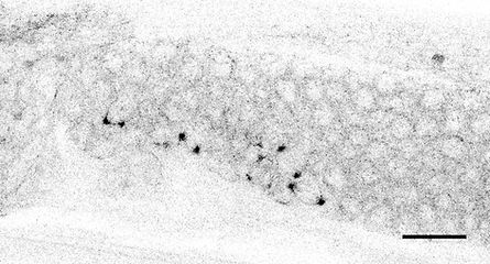
The C. elegans adult hermaphrodite gonad houses two pools of totipotent adult germline stem cells (GSCs). Similar to mammalian stem cells, adult GSCs self-renew, are maintained by proximity-based niche interactions and are responsive to environmental conditions. Unlike other adult stem cells, GSC development is well defined and GSCs offer unparalleled accessibility for live-imaging studies. GSCs are derived from a single precursor cell, which itself is specified early during embryonic development. Because C. elegans embryogenesis is invariant and all cell lineages have been fully mapped, we can also perform live imaging of GSCs precursors (germline blastomeres) to determine how certain traits emerge during development.
Research in the lab is focused around three primary questions:
How is spindle assembly checkpoint strength modified by cell size and cell lineage during embryogenesis? (more...)
How is mitotic fidelity maintained when GSCs divide under different physiological or environmental conditions? (more...)
How is GSC spindle orientation and cytokinesis adapted to maintain germline tissue organization? (more...)
This work is supported by funds from:


